Ventricular Septal Defect (VSD)

Ventricular Septal Defect (VSD)

 Ventricular Septal Defect (VSD) is a congenital disease, which affects the septum – the wall in between the left and right ventricles of the heart. This opening enables oxygen, rich blood, to be permitted to mix with oxygen-poor blood in the right ventricle thereby enhancing the amount of blood flow in the lungs. VSD is the second most frequently occurring congenital heart disease, and the defect can be of an assortment of sizes. While small-sized VSDs are believed to close spontaneously during infancy, larger-sized VSDs often need medical interventional treatment or surgical repairage.

The symptoms of VSD also depend with the size of the defect and the degree of blood flow. Common symptoms include:

  • Shortness of Breath: Shortness of breath or discomfort when breathing, or sudden breathlessness with feeds in babies under a month.
  • Rapid Breathing: A high level of respiration whereby the breathing rate goes above the normal rate and may include grunting and wheezing.
  • Poor Growth: Poor weight gain, failure to thrive in infancy as a result of the rising workload of the heart.
  • Fatigue: Exhaustible during activities, this may be so especially in children or infants.
  • Frequent Respiratory Infections: Pneumonia for example one is likely to catch an infection when his or her immune system is weak.
  • Heart Murmur: A high pitched, blowing and rushing sound, often detected during the physical assessment that occurs due to the blood flow through the defect.
  • Breathlessness, more specifically exercised-induced breathlessness
  • Fatigue
  • Leg and/or foot or lower abdominal enlargement
  • Palpitations or extrasystoles,MouseEvent: Other ERRs
  • Stroke
  • Heart murmur, a whooshing sound, and that, when the doctor puts the stethoscope on the chest.
Symptoms Collapse Symptoms
  • Infants Show Signs of Poor Growth: It refers to an inability to grow properly, or otherwise if an infant is not taking weight or growing as expected.
  • Breathing Difficulties: If your baby experiences any of the following, then this could be a sign of RSV: constant coughing, ongoing shortness of breath, noticeable increase in breathing rate or breathing hard while feeding.
  • Fatigue or Lack of Energy: If a child looks so much exhausted during exercise that he or she should not look exhausted.
  • Frequent Respiratory Issues: Repeated cases of lower respiratory tract infection or symptoms of breathing difficulty for example coughing and wheezing.
  • Heart Murmur Detected: If there is a heart murmur during the physical examination of the patient, then there is a need for more tests.
When to Go for a Medical Check-up Collapse When to Go for a Medical Check-up

 VSD is an acquired pathology, which in most cases is diagnosed in childhood. It occurs due to:

  • Incomplete Development of the Heart Septum: In development of the fetal, the septum is incompletely developed so that there is an opening between the ventricles.
  • Genetic Factors: VSD is known to be related with other genetic disorders or chromosomal impairments including down syndrome.
  • Environmental Factors: It has also been viewed that conditions like maternal illness, use of medication or any other substance during pregnancy can lead to congenital heart defects including VSD.
Causes Collapse Causes

 Diagnosis of VSD typically involves the following steps:


  • Physical Examination: A health care provider may hear a murmur during an ordinary physical assessment and make a referral for additional assessment.
  • Echocardiogram: An echocardiogram that creates images of the heart and can determine the size and location or the VSD and its effect on the heart.
  • Chest X-ray: This may be employed to look for features of an enlarged heart or raised blood vessels in the lungs.
  • Electrocardiogram (ECG): An accuracy of the heart muscles activity and a capability of an indication of an enlarged heart, or other irregularities.
  • Cardiac Catheterization: Sometimes, a thin flexible tube called catheter is introduced through the blood vessels to enter the heart and record pressures within the heart chambers as well as to have a better view of the defect.
Diagnosis Collapse Diagnosis

 Diagnosis of VSD typically involves the following steps:

  • Physical Examination: A health care provider may hear a murmur during an ordinary physical assessment and make a referral for additional assessment.
  • Echocardiogram: An echocardiogram that creates images of the heart and can determine the size and location or the VSD and its effect on the heart.
  • Chest X-ray: This may be employed to look for features of an enlarged heart or raised blood vessels in the lungs.
  • Electrocardiogram (ECG): An accuracy of the heart muscles activity and a capability of an indication of an enlarged heart, or other irregularities.
  • Cardiac Catheterization: Sometimes, a thin flexible tube called catheter is introduced through the blood vessels to enter the heart and record pressures within the heart chambers as well as to have a better view of the defect.
Treatment Collapse Treatment

efficient and better care for you.

We prioritize delivering care that is both efficient and effective, ensuring you receive the best possible treatment with minimal wait times. Our focus is on providing high-quality, personalized care that enhances your overall well-being and health outcomes.
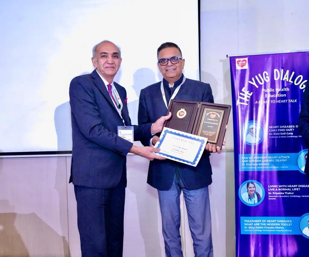
 

Our Timings

Monday to Saturday: 9am - 5pm
Sunday: Closed